Capabilities in Autoimmune and
Inflammation Platform
Capabilities in Autoimmune and Inflammation Platform
Autoimmune and Inflammation Animal Models Overview
Autoimmune Disease Models
Experimental Autoimmune Encephalomyelitis (EAE)

MOG35-55/MOG1-125-induced EAE in mice

MBP-induced EAE in rats

PLP139-151-induced EAE in mice

Rheumatoid Arthritis (RA)

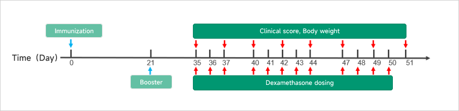
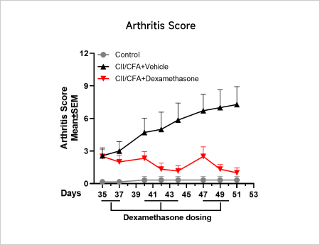
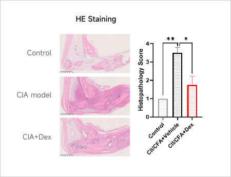
Collagen-induced arthritis (CIA) in mice and rats

Collagen antibody-induced arthritis (CAIA) in mice

Adjuvant-induced arthritis (AIA) in rats

Systemic Lupus Erythematosus (SLE)

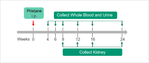
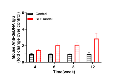
Pristane-induced lupus in mice

MRL/lpr mice lupus model

Inflammatory Bowel Disease (IBD)

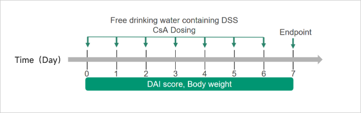
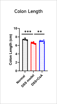
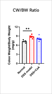
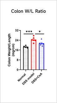
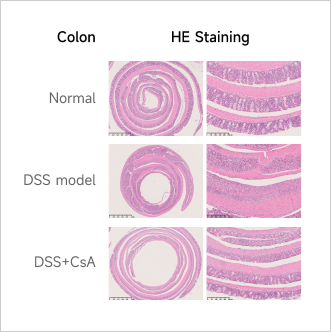
DSS-induced ulcerative colitis in mice

TNBS-induced crohn's disease in mice

IL-10 Knockout spontaneous colitis model

CD4+CD45RBhigh T cell-induced colitis in mice

Allergic Disease Models
Atopic Dermatitis (AD)

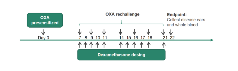
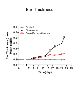
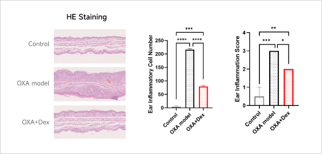
Oxazolone-induced atopic dermatitis in mice

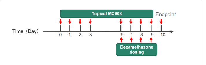
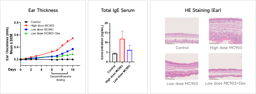
MC903-induced dermatitis in mice

DNCB-induced dermatitis in mice and rats

FITC-induced dermatitis in mice

Psoriasis

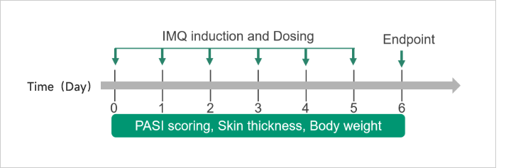
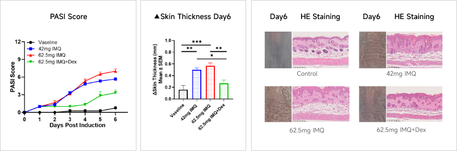
Imiquimod-induced psoriasis in mice

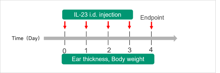
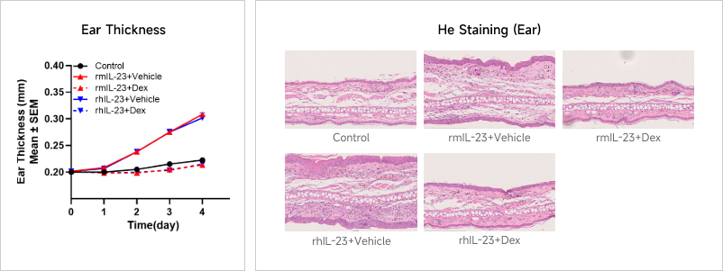
IL-23-induced ear epidermal hyperplasia in mice

Asthma

OVA-induced asthma in mice

HDM-induced asthma in mice

Delayed Type Hypersensitivity (DTH)

Oxazolone-induced DTH in mice

KLH-induced DTH in mice

Fibrosis Models
Liver Fibrosis

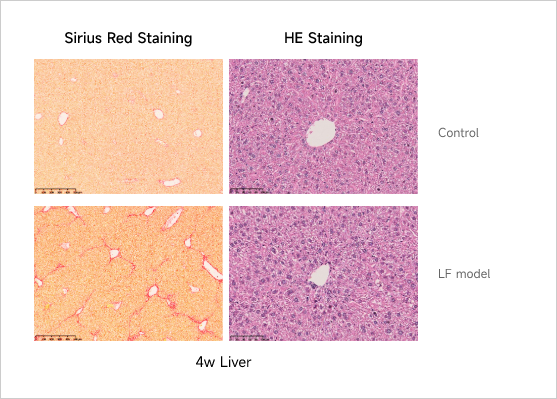
CCl4-induced liver fibrosis in mice and rats

DMN-induced liver fibrosis in mice and rats

TAA-induced liver fibrosis in mice and rats

Bile Duct Ligation (BDL) model in mice and rats

Pulmonary Fibrosis

Bleomycin-induced pulmonary fibrosis in mice

Kidney Fibrosis

Folic acid-induced kidney fibrosis in mice

Short Term Inflammation Models
LPS-induced Acute Inflammation Model
APAP-induced Acute Liver Injury Model
T cell Dependent Antibody Response (TDAR) Model

KLH-induced TDAR in mice

Eosinophilic Gastroenteritis (EG)

OVA-induced eosinophilic gastroenteritis in mice

Air Pouch Model

Carrageenan-induced air pouch in mice and rats

rhIL-13-induced air pouch in mice

GvHD Models
Graft-versus-Host Disease (GvHD)

GvHD induced by X-ray irradiation in mice

GvHD without irradiation in rats

  • Rheumatoid Arthritis (RA)
     
    Case Study: Collagen-induced Arthritis (CIA) Model
    Animals: DBA/1, 7-8 weeks, male
    Modeling reagent: Bovine CII + CFA emulsion
    Modeling method: Immunization: day 0, i.d., tail Booster: day 21
    Readouts
    Clinical observation: Limb score, BW
    Histopathology: H&E、Synovial inflammation、Bone Erosion
    Clinical and Histological Assessment in Collagen-induced CIA Model
  • Systemic Lupus Erythematosus (SLE)
     
    Case Study: Pristane-induced SLE Model
    Experimental animals: Balb/c, 6-8 weeks, female
    Modeling reagent: Pristane
    Modeling method: 0.5ml, i.p., once
    Readouts
    Clinical observation:  Body weight
    Histopathology:  H&E、IHC or IF
    Urine chemistry analysis:  Creatinine, UTP, mALB, UREA
    Cellular and molecular levels:  Anti-dsDNA IgG 、Cytokine level 、FACS
    Anti-dsDNA Analysis in Pristane-induced SLE Model
  • Inflammatory Bowel Disease (IBD)
     
    Case Study: DSS-induced Acute Ulcerative Colitis Model
    Experimental animals: C57BL/6, 7 weeks, female
    Modeling reagent: Dextran Sulfate Sodium (DSS)
    Modeling method: Free drinking water containing DSS for consecutive 7 days
    Readouts
    Clinical observation: DAI score, BW
    Histopathology: Colon H&E staining
    Molecular levels: Cytokine level
    Clinical and Histological Assessment in DSS-induced UC Model
  • Atopic Dermatitis (AD)
     
    Case Study: Oxazolone-induced Atopic Dermatitis (AD) Model
    Experimental animals: Balb/c, 6-8 weeks, female
    Modeling reagent: Oxazolone (OXA)
    Modeling method: Sensitization: day 0 on back
    Rechallenge: day7-21 on right ear, 7 times
    Readouts
    Clinical observation: Ear thickness, BW
    Histopathology: H&E、Immune infiltration
    Cellular and molecular levels: FACS analysis、Cytokine level、Total IgE
    Clinical and Histological Assessment in OXA-induced AD Model
    Case Study: MC903-induced Atopic Dermatitis (AD) Model
    Experimental animals: C57BL/6, 7-8 weeks, female
    Modeling reagent: MC903
    Modeling method: Topically, on ear, 8 times
    Readouts
    Clinical observation: Ear thickness, BW
    Histopathology: H&E staining
    Cellular and molecular levels: Cytokine level、Total IgE
    Clinical and Histological Assessment in MC903-induced AD Model
  • Psoriasis
     
    Case Study: IMQ-induced Psoriasis Model
    Experimental animals: C57BL/6, 7 weeks, female
    Modeling reagent: Imiquimod (IMQ) cream
    Modeling method: Topically applicated on dorsal for consecutive 6 days
    Readouts
    Clinical observation: Skin thickness, PASI score, BW
    Histopathology: H&E staining
    Cellular and molecular levels: Cytokine level、FACS
    Clinical and Histological Assessment in IMQ-induced Psoriasis Model
    Case Study: IL-23-induced Ear Epidermal Hyperplasia Model
    Experimental animals: C57BL/6, 7 weeks, female
    Modeling reagent: Recombinant mouse IL-23 and human IL-23
    Modeling method: Intradermal injection, ear, 4 times
    Readouts
    Clinical observation: Ear thickness, BW
    Histopathology: H&E staining
    Cellular and molecular levels: Cytokine level、FACS
    Clinical and Histological Assessment in IL-23-induced Ear Epidermal Hyperplasia Model
  • Asthma
     
    Case Study: OVA-induced Asthma Model
    Experimental animals:Balb/c, 6-8 weeks, female
    Modeling reagent:Ovalbumin (OVA) + Al(OH)3
    Modeling method:Sensitization: i.p., on day 0, 7, 14 Rechallenge: Intranasal, on day21-25
    Readouts
    Clinical observation:Body weight, Health condition
    Histopathology:H&E, PAS Staining、IHC
    Cellular and molecular levels:FACS analysis、Cytokine level、Total IgE, OVA specific IgE
    Histological Assessment in OVA-induced Asthma Model

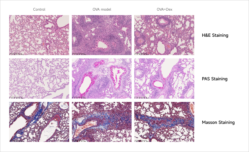
    After OVA induction, there is a significant infiltration of inflammatory cells in the lung tissue, and the structure of the airway is disordered, and the normal structure of the bronchi and alveoli is damaged.

    There is a significant increase in the number of goblet cells in the airway epithelium. The PAS staining highlights a large amount of mucus secretion.

    Due to the experimental cycle, pulmonary fibrosis is not obvious after OVA induction.

    Dexamethasone treatment ameliorates the pathological changes, including reducing goblet cell hyperplasia, decreasing mucus secretion, alleviating inflammatory cell infiltration, and improving airway and tissue architectural integrity.

    Total IgE and Cytokine Analyses in OVA-induced Asthma Model

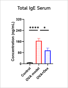
    A significant increase in the level of total IgE can be observed in serum after OVA induction.

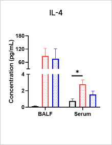
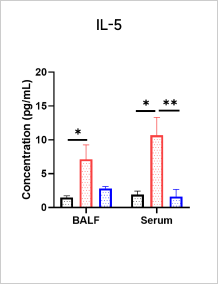
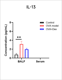
    After OVA induction, the expression of Th2-type cytokines (IL - 4, IL - 5, IL - 13) is up-regulated and Dexamethasone treatment can reduce the levels of IL - 4, IL - 5, and IL-13 and total IgE.

  • Liver Fibrosis
     
    Case Study: CCl4-induced Liver Fibrosis Model
    Experimental animals:C57BL/6, 6-8 weeks, male
    Modeling reagent:CCl4/Olive oil
    Modeling method:BIW, i.p., 8 weeks
    Readouts
    Clinical observation:Body weight, Health condition
    Histopathology:H&E staining, Masson or SR staining、IHC (α-SMA)
    Serum biochemical:ALT, AST, TBA, ALB, ALP
    Histopathological Analysis in CCl4-induced Liver Fibrosis Model
  • LPS-induced Acute Inflammation
     
    Case Study: LPS-Induced Acute Inflammation Model
    Objective:To evaluate the effect of Azathioprine and WAY-171318 in LPS-induced acute inflammation model
    Animal:C57BL/6, 6-8 weeks, female
    Modeling reagent:LPS + D-Galactosamine
    Modeling method:
    Cytokine Analyses iN LPS-Induced Inflammation Model
For more information about

Pharmacology & DMPK”, please contact our team.

Contact Us
Online Message
After receiving your message, our professional team will contact you shortly.
  • Area of Interest

    CRO Drug R&D Services

    CDMO R&D and Production Services

    EFS Investment&Incubation Business

    Media Cooperation

    Investor Relations

    Other

    How Did You Hear About Us?

    Search Engine (Google, Bing, Yahoo, etc)

    Conferences/Events

    Social Media

    LinkedIn

    Word of Mouth

    Other